Thursday, 30 January 2020

Infective Endocarditis: Types

Defination

In simple words, It’s a microbial infection of heart valves or lining of endocardium.
The microbial organism can be bacteria or parasite or fungus or rickettsia or chlamydia.

Let’s check out different types!

  1. Subacute Bacterial Endocarditis 
  2. Acute Infective Endocarditis 
  3. Post Operative Endocarditis 
  4. Right Sided Endocarditis 

What’s Subacute Endocarditis? 

  • It is caused by relevantly low virulence organisms like streptococcus viridan
  • It mainly effects 
    • damaged heart valves
    • MacCallum plaque which is irregular thickness usually found in the left atrium in patients with rheumatic fever
    • Also, Low pressure areas of heart 
  • What are the Characteristics Features? It’s simple 4 points 🙂 
    • Formation of vegetation 
    • Emboli formation 
    • Mycotic aneurism 
    • Valvular Regurgitation

What is Acute Infective Endocarditis?

  • It is caused by high virulence organism like staph. aureus 
  • It effects both normal and damaged valves
  • Doc, you need to be careful cause clinical course can be fatal if untreated within 6 weeks!!
  • What are the Clinical features? How is it different from subacute?
    • Valve destruction is greater 
    • Abscess formation is common
    • Valve cusp perforation can also occur

What is Post-operative Endocarditis? 

  • As the name indicates, during cardiac surgery, the patient develops infective endocarditis 
  • What is a prosthetic valve? It’s an artificial valve replacing the mitral or aortic valve 
  • It mainly affects the prosthetic valves, especially aortic valve 
  • Source of infection- staph epidermis 
  • Generally, within 3-60 days of a health care facility admission, the nosocomial infections will cause endocarditis
  • It accounts for 20% of Infectious Endocarditis. So, provide clean facilities in hospital, doc.
  • Typically, it is associated with invasive procedures like dental procedures and intravenous access.

What is Right-sided Endocarditis?

  • Who is mainly affected? Intravenous drug addicts 
  • It is caused when they share a syringe with other people during drug abuse
  • Source of infection: staph aureus and candida present on the surface of the skin
  • So, the microorganisms enters into the body through veins during drug abuse.
  • The right side of the heart is affected, especially tricuspid valve.
  • Larger bloodborne particulate matter in IV drug abusers typically deposits on the tricuspid valve.
  • Remember, tricuspid valve is rarely involved in other causes of Bacterial Endocarditis
  • Generally, the clinical course will be subacute or chronic or insidius.
Tuesday, 28 January 2020

Infective Endocarditis- Pathophysiology

What are the sites where the infection can occur? Cause these are areas of the nidus where the infection can occur! 
  • Normal cardiac endothelium 
  • Damaged valves
  • Mitral valve and aortic valve are most likely to be involved
  • Surgically constructed AV shunts 
  • Prosthetic valves
Imagine, a breach in the endothelium caused by
  • Turbulent flow (e.g. valvular stenosis or valvular regurgitation) or 
  • Intravascular device-related injury (e.g. catheters)
After the breach, the platelets and WBC’s aggregate near injured endothelium like soldiers and give rise to the thrombus.
If dental procedures or any surgery takes place after thrombi formation, chances of transitent bacterium to colonize the thrombi is more.
Hence, immune system will be form fibrin meshwork on bacteria to seal it.
So, thrombus + bacteria + fibrin = is called vegetation 
When vegetation is dislodged, it may go anywhere in the body. 
  • It can form embolus
  • Since, it contains bacteria, it’s septicaemia in nature 
  • It contains foreign particles too, hence immunogenic in nature

Sunday, 12 January 2020

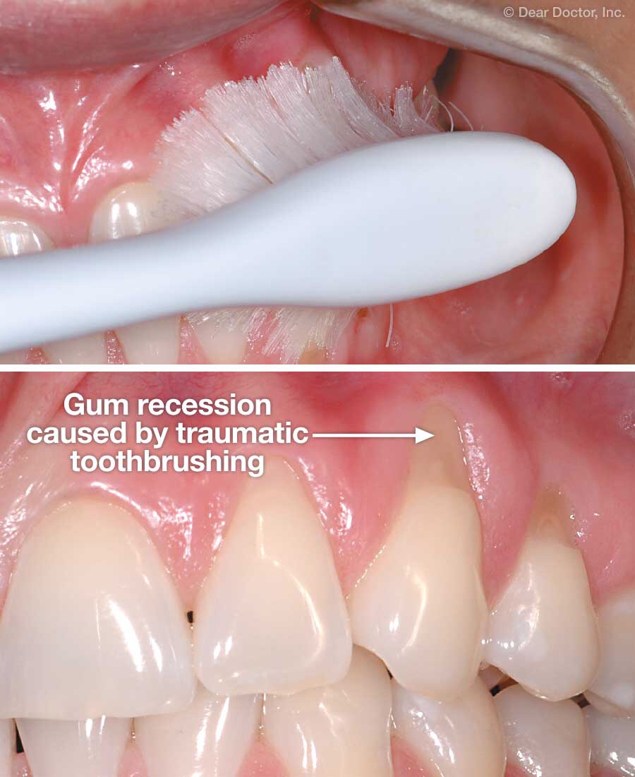
Abrasion

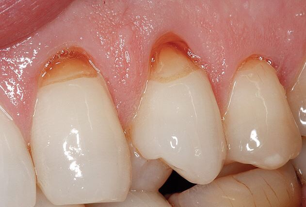
In simple words, Abrasion is loss of tooth structure due to foreign substances , like heavy brushing, hard bristels. Abrasion occours in the cervical region of tooth

Etiology

  • Faulty oral hygiene practice 
    • Horizontal brushing 
    • Excessive forces
  • Quality of toothbrush 
  • pH and amount of dentifrice used 
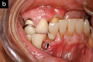
  • Ill-fitting clasps of partial dentures cause localised abrasion lesions
  • Fiction from toothpicks and interproximal brushes
  • Tobacco Chewing

Treatment

We need to take careful consideration of aetiology and progression of the condition. That means, correct diagnosis is the prerequisite for the management of the lesion.
  • If the lesion is localized and not interfering with the physiological function of the stomatognathic system = It may be restored 
  • If the abrasion is generalized and substantial = the habit should be discontinued and controlled
  • If teeth are sensitive = use Flouride application 
  • If it’s class V lesion = Restoration with GIC
  • If lesion involves a none conscious area in the posterior teeth = use metallic restorationon

Attrition

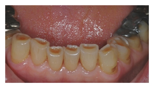
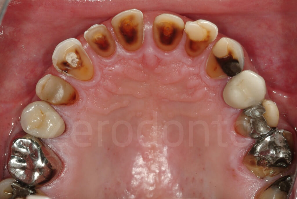
In simple words, Attrition is the loss of tooth structure occlusally due to excessive forces by the occluding teeth, grinding of teeth, deep bite

Aetiology

  • Age 
  • Hard and Abrasive diet 
  • Chewing Habits 
  • Opposing porcelain crowns
  • Lack of posterior support- cause of anterior teeth attrition
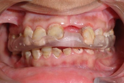
  • Bruxism and Clenching
Clinical Manifestations
Occluding surface attrition causes small polished facet on a cusp tip or ridge or incisal edge 
In severe cases, reverse cusp in place of cusp tips and inclined planes. This causes dentinal exposure and loss of vertical dimension of teeth.
Precipating Factors
  • Deficient masticatory capabilities of teeth
  • Cheek biting
  • Caries- because of the increased vulnerability of exposed dentinal surfaces 
  • TMJ Problems
  • Jutted out appearance of restoration and flattening of surrounding occlusal surface. %
Treatment : Pathologic
  • Advice the patient to wear splints 
  • To manage localised anterior teeth wear: use dahl technique
  • The more traditional way of dealing with a full arch or full mouth rehabilitation is to use conventional crowns eg, onlays of a full crown


Different Types of Proteoglycan Sulfates

Chondroitin sulfates are the most prominent proteoglycans in cartilage and bone. Keratin sulfates are most prominent in the cornea (type...